15 + Corona Radiata Infarct Radiology HD Resolutions. Evidence from subcortical small infarcts suggests that motor fibers are somatotopically arranged in the human corona radiata. "Old infarct in left centrum semi ovals and frontal corona radiata?" Answered by Dr. Inferiorly these tracts converge as the internal capsule.

21 + Corona Radiata Infarct Radiology High Quality Images
Appear as wedge-shaped, cortically based, hypodense areas.

Acute Brain Infarction – Something About Radiology – Just ...
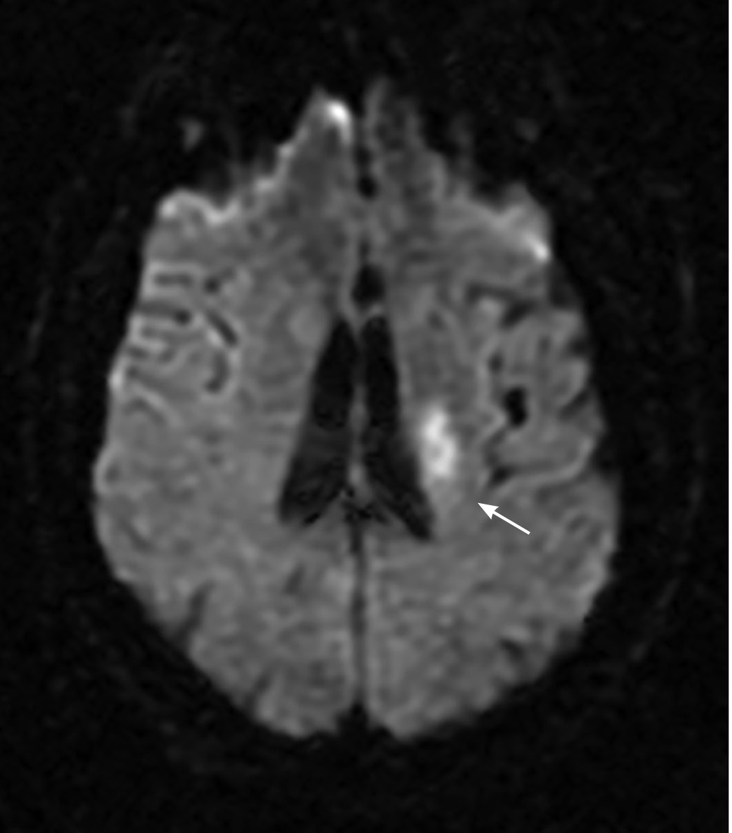
Transient Ischemic Attack | 2019-02-05 | Relias Media ...
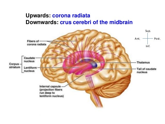
Internal Capsule-Anatomy

Acute infarction | Image | Radiopaedia.org
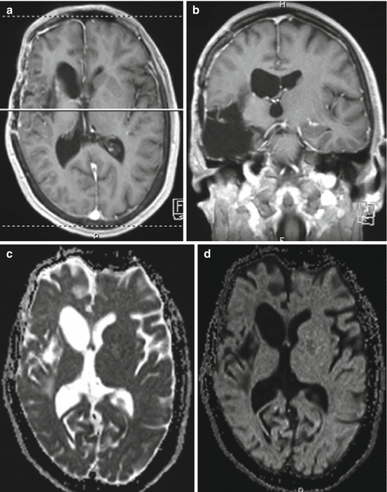
Arteries and Veins of the Sylvian Fissure and Insula ...

ECR 2014 / C-2095 / Approach to Non-Traumatic ...

The infarct location predicts progressive motor deficits ...

Ipsilateral Hemiparesis Caused by a Corona Radiata Infarct ...
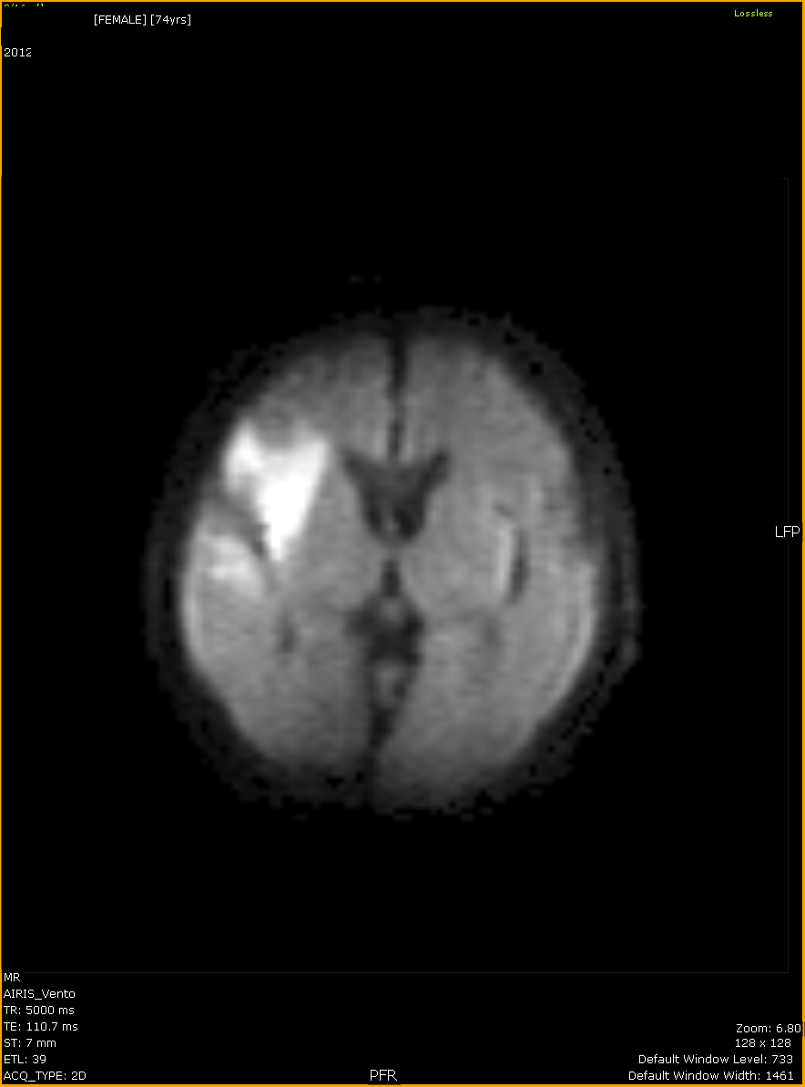
Acute Brain Infarction – Something About Radiology – Just ...

Central nervous system manifestations in human ...

Small acute left MCA infarct — Clinical MRI
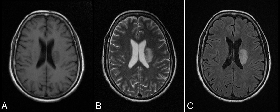
Early detection of secondary damage in ipsilateral ...
:max_bytes(150000):strip_icc()/doctor-viewing-a-series-of-mri--magnetic-resonance-imaging--brain-scans-on-a-screen-592232673-5b4df38d46e0fb005b1151b8.jpg)
Damage to the Corona Radiata After Stroke
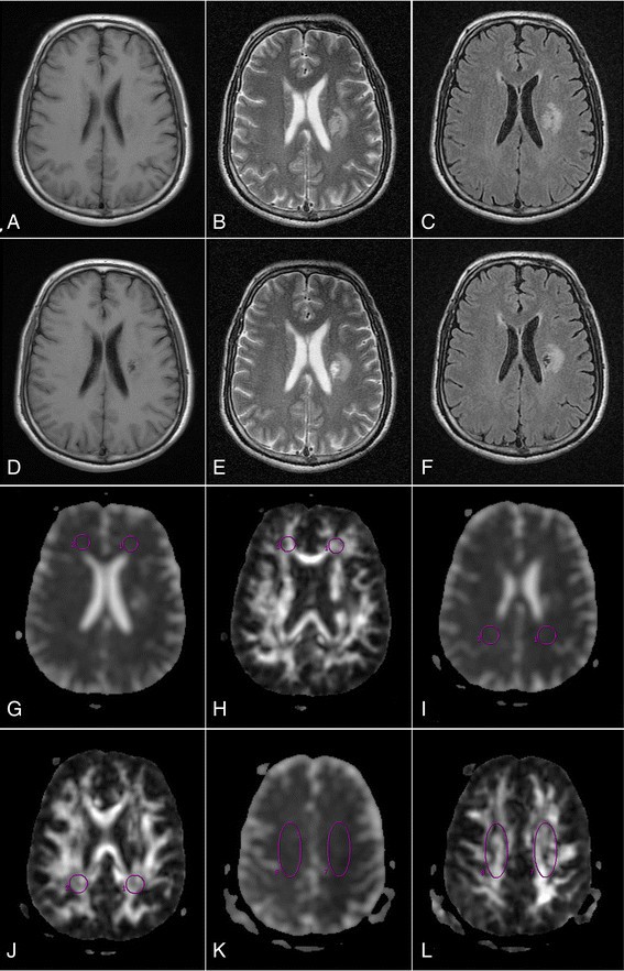
Secondary damage in left-sided frontal white matter ...
Dr Balaji Anvekar FRCR: Ischemic stroke and Vascular ...
15 + Corona Radiata Infarct Radiology HD ResolutionsAbstract: Objectives:Diffusion tensor image tracography (DTT) could be useful for exploration of the state of the corticospinal tract at the subcortical white matter level. Find out information about corona radiata. In neuroanatomy, the corona radiata is a white matter sheet that continues ventrally as the internal capsule and dorsally as the semioval center.

