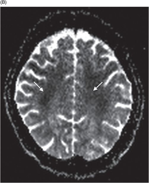
15 + Centrum Semiovale And Corona Radiata Radiology Desktop Wallpaper. Related pathology. centrum semiovale (along with the basal ganglia) are a. It also contains commissural, projection, and association fibers.

21 + Centrum Semiovale And Corona Radiata Radiology High Quality Images
In neuroanatomy, the corona radiata is a white matter sheet that continues ventrally as the internal capsule and dorsally as the centrum semiovale.

ECR 2013 / C-1430 / Diffusion-Weighted MR Imaging ...

Corona Radiata Brain - Eyebrows Idea

Corona Radiata Brain Ct Anatomy - Eyebrows Idea
What is the function of the right centrum semiovale? - Quora

65 | Radiology Key

Infarction: Infarct Corona Radiata

ECR 2010 / C-3395 / Conventional magnetic resonance and ...

Multiple sclerosis | Radiology Case | Radiopaedia.org

Diffusion Tensor Imaging to Predict Long-term Outcome ...

Centrum Semiovale Anatomy - HealthCop.com - Your Health Cop

ECR 2010 / C-2613 / Prediction of a large diffusion ...

Multiple Sclerosis Presenting as Bell Palsy | Neurology

Anatomy Corona Radiata Ct - Eyebrows Idea

Petro-clival meningioma | Radiology Case | Radiopaedia.org

Normal Anatomy | Radiology Key
15 + Centrum Semiovale And Corona Radiata Radiology High Quality ImagesCentrum semiovale information including symptoms, causes, diseases, symptoms, treatments, and other medical and health issues. Inferiorly these tracts converge as the internal capsule. The centrum semiovale is the white matter deep to the gray matter on the surface of the brain and has an ovular shape.

