15 + Corona Radiata Vs Centrum Semiovale Radiology Background Images. Superiorly they are continuous with the centrum semiovale. This sheet of both ascending and descending axons carries most of the neural traffic from and to the cerebral cortex.

21 + Corona Radiata Vs Centrum Semiovale Radiology HD Resolutions
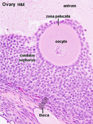
The corona radiata is an outer layer of follicular (granulosa) cells that form around a developing oocyte in the ovary and remain with it upon ovulation.

31. The anatomy, histology and development of the ovary ...


File:Schematic reconstruction of association, projection ...

ShareMyRadiology 放射线学: October 2012
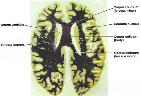
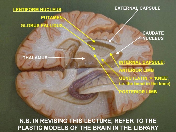
Plate 17.357 Caudate Nucleus

Lesional demyelinations of the central nervous system ...

The Radiology Assistant : Solitary pulmonary nodule ...

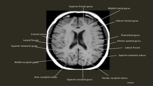
인체여행 - A journey into the human body : Parietal gyri

Brain CT - NeurologyNeeds.com
Table #10
Anatomy of Motor system2
Fertilization - Embryology
Ns9. Motor Pathways. Compressed File
Anatomy
Centrum Semiovale Ct Brain Anatomy Pictures to Pin on ...
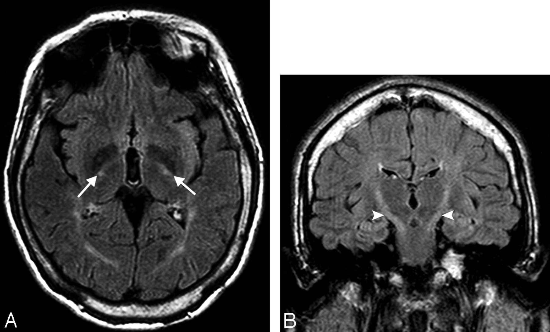
MR Imaging Findings in Autosomal Recessive Hereditary ...
15 + Corona Radiata Vs Centrum Semiovale Radiology Background ImagesThe corona radiata is associated with. Superiorly they are continuous with the centrum semiovale. On the upper parts of the fibers, they appear continuous with coronal radiata.