15 + Corona Radiata And Centrum Semiovale Mri HD Resolutions. The common central mass of white matter with an oval appearance in horizontal sections of the brain is termed the centrum semiovale. In neuroanatomy, the corona radiata is a white matter sheet that continues ventrally as the internal capsule and dorsally as the centrum semiovale.
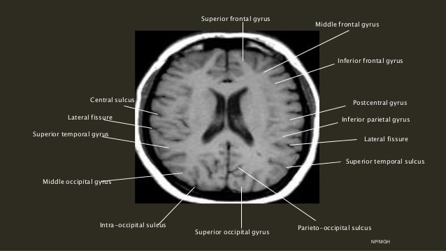
21 + Corona Radiata And Centrum Semiovale Mri Background Images
Lesions affecting the frontal-subcortical circuits (e.g. pallidum, corona radiata or centrum semiovale) were more frequent in patients with ED than in.

Corona Radiata Brain Ct Anatomy - Eyebrows Idea

Internal watershed zone cerebral infarction | Image ...

Caudate Infarct

ECR 2011 / C-1606 / Multimodality Neuroimaging in Sickle ...

View Image

Neurointerventional Treatment of Acute Stroke in 2015 at ...

Illustrative MRI of the brain and cervical spinal cord ...
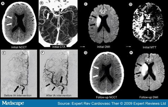
corona radiata stroke

Ipsilateral Hemiparesis Caused by a Corona Radiata Infarct ...

Multiple sclerosis | Radiology Case | Radiopaedia.org

Cerebrum ii
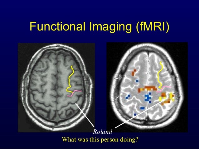
Anatomy of Motor system2
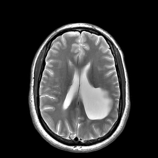
Porencephaly | Eurorad

ECR 2010 / C-3395 / Conventional magnetic resonance and ...
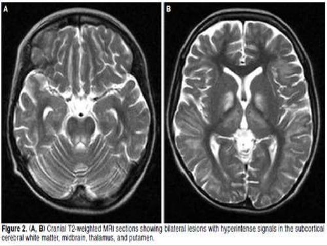
Mri evaluation of pediatric white matter lesions
15 + Corona Radiata And Centrum Semiovale Mri High Quality ImagesAxial FLAIR MRI through the corona radiata and centrum semiovale, respectively. Inferiorly these tracts converge as the internal capsule. Hi, This report means there is acute infaction of internal capsule,corona radiata and centrum semiovale.

